A short History of the development of Ultrasound in Obstetrics and Gynecology
Dr. Joseph Woo
As early as 1826, Jean-Daniel Colladon, a Swiss physicist, had successfully used an underwater bell to determine the speed of sound in the waters of Lake Geneva. In the later part of the 1800s, physicists were working towards defining the fundamental physics of sound vibrations (waves), transmission, propagation and refraction.
The real breakthrough in the evolution of high frequency echo-sounding techniques came when the piezo-electric effect in certain crystals was discovered by Pierre Curie and his brother Jacques Curie in Paris, France in 1880. They observed that an electric potential would be produced when mechanical pressure was exerted on a quartz crystal such as the Rochelle salt (sodium potassium tartrate tetrahydrate). The reciprocal behavior of achieving a mechanical stress in response to a voltage difference was mathematically deduced from thermodynamic principles by physicist Gabriel Lippman in 1881, and which was quickly verified by the Curie brothers. It was then possible for the generation and reception of 'ultrasound' that are in the frequency range of millions of cycles per second (megahertz) which could be employed in echo sounding devices. Further research and development in piezo-electricity soon followed.
Langevin's hydrophones had formed the basis of the development of naval pulse-echo sonar in the following years. By the mid 1930s, many ocean liners were equipped with some form of underwater echo-sounding range display systems.
Such radar display systems had been the direct precursors of subsequent 2-dimensional sonars and medical ultrasonic systems that appeared in the late 1940s. Books such as the "Principles of Radar" published by the Massachusetts Institute of Technology (M I T) Radar school staff in 1944 detailed the techniques of oscilloscopic data presentation which were employed in medical ultrasonic research later on (see below). Two other engineering advances probably had also influenced significantly the development of the sonar, in terms of the much needed data aqusition capabilities: the first digital computer (the Electronic Numerical Integrator and Computer -- the ENIAC) constructed at the University of Pennsylvania in 1945, and the invention of the point-contact transister in 1947 at AT & T's Bell Laboratories.
The concept of ultrasonic metal flaw detection was first suggested by Soviet scientist Sergei Y Sokolov in 1928 at the Electrotechnical Institute of Leningrad. He showed that a transmission technique could be used to detect metal flaws by the variations in ultrasionic energy transmitted across the metal. The resolution was however poor. He suggested subsequently at a later date that a reflection method may be practical.
The equipment suggested by Sokolov which could generate very short pulses necessary to measure the brief propagation time of their returning echoes was not available until the 1940s. Early pioneers of such reflective metal flaw detecting devices were Floyd A Firestone at the University of Michigan, and Donald Sproule in England. Firestone produced his patented "supersonic reflectoscope" in 1941 (US-Patent 2 280 226 "Flaw Detecting Device and Measuring Instrument", April 21, 1942). Because of the war, the reflectoscope was not formally published until 1945. Messrs. Kelvin and Hughes® in England, where Sproule was working, had also produced one of the earliest pulse-echo metal flaw detectors, the M1. Josef and Herbert Krautkrämer produced their first German version in Köln in 1949 followed by equipment from Karl Deutsch in Wuppertal. These were followed by other versions from Siemens® in Erlangen, KretzTechnik AG in Austria, Ultrasonique in France and Mitsubishi in Japan. In 1949, Benson Carlin at M I T, and later at Sperry Products, published "Ultrasonics", the first book on the subject in the English language.
[Home] [ Part 1 ] [ Part 2 ] [ Part 3 ] [ Site Index ]
site search by
freefind
advanced
 he story of the development of ultrasound applications in medicine should probably start with the history of measuring distance under water using sound waves. The term SONAR refers to Sound Navigation and Ranging. Ultrasound scanners can be regarded as a form of 'medical' Sonar.
 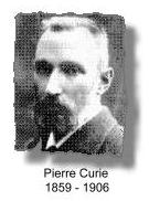 One of them was Lord Rayleigh in England whose famous treatise "the Theory of Sound" published in 1877 first described sound wave as a mathematical equation, forming the basis of future practical work in acoustics. As for high frequency 'ultrasound', Lazzaro Spallanzani, an Italian biologist, could be credited for it's discovery when he demonstrated in 1794 the ability of bats navigating accurately in the dark was through echo reflection from high frequency inaudible sound. Very high frequency sound waves above the limit of human hearing were generated by English scientist Francis Galton in 1876, through his invention, the Galton whistle.
  Underwater sonar detection systems were developed for the purpose of underwater navigation by submarines in World war I and in particular after the Titanic sank in 1912. Alexander Belm in Vienna, described an underwater echo-sounding device in the same year. The first patent for an underwater echo ranging sonar was filed at the British Patent Office by English metereologist Lewis Richardson, one month after the sinking of the Titanic. The first working sonar system was designed and built in the United States by Canadian Reginald Fessenden in 1914. The Fessenden sonar was an electromagnetic moving-coil oscillator that emitted a low-frequency noise and then switched to a receiver to listen for echoes. It was able to detect an iceberg underwater from 2 miles away, although with the low frequency, it could not precisely resolve its direction.
 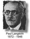 The turn of the century also saw the invention of the Diode and the Triode, allowing powerful electronic amplifications necessary for developments in ultrasonic instruments. Powerful high frequency ultrasonic echo-sounding device was developed by emminent French physicist Paul Langévin and Russian scientist Constantin Chilowsky, then residing in France. Patents were filed in France and the United States. They called their device the 'hydrophone'. The transducer of the hydrophone consisted of a mosaic of thin quartz crystals glued between two steel plates with a resonant frequency of 150 KHz. Between 1915 and 1918 the hydrophone was further improved in classified research activities and was deployed extensively in the surveillance of German U-boats and submarines. The first known sinking of a submarine detected by hydrophone occurred in the Atlantic during World War I in April,1916.
 In another development, the first successful radio range-finding experiment occurred in 1924, when British physicist Edward Appleton used radio echoes to determine the height of the ionosphere. The first practical RADAR system (Radio Detection and Ranging, and using electromagnetic waves rather than ultrasonics) was produced in 1935 by another British physicist Robert Watson-Watt, and by 1939 England had established a chain of radar stations along its south and east coasts to detect aggressors in the air or on the sea. World war II saw rapid developments and refinements in the naval and military radar by researchers in the United States.
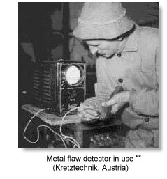 Yet another parallel and equally important development in ultrasonics which had started in the 1930's was the construction of pulse-echo ultrasonic metal flaw detectors, particularly relevant at that time was the check on the integrity of metal hulls of large ships and the armour plates of battle tanks.
The underwater SONAR, the RADAR and the ultrasonic Metal Flaw Detector were each, in their unique ways, a precursor of medical ultrasonic equipments. The modern ultrasound scanner embraces the concepts and science of all these modalities.
 The early development of ultrasonics is summarised here.
 Readers are also referred to an article by Dr William O'Brien Jr., which also looks at the early history of the developments of ultrasonics.^
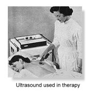
 he use of Ultrasonics in the field of medicine had nonetheless started initially with it's applications in therapy rather than diagnosis, utilising it's heating and disruptive effects on animal tissues. The destructive ability of high intensity ultrasound had been recognised in the 1920s from the time of Langévin when he noted destruction of school of fishes in the sea and pain induced in the hand when placed in a water tank insonated with high intensity ultrasound; and from the seminal work in the 1930s from Robert Wood, Newton Harvey and Alfred Loomis in New York and R Pohlman in Erlangen, Germany.
High intensity ultrasound progressively evolved to become a neuro-surgical tool. William Fry at the University of Illinois and Russell Meyers at the University of Iowa performed craniotomies and used ultrasound to destroy parts of the basal ganglia in patients with Parkinsonism. Peter Lindstrom in San Francisco reported ablation of frontal lobe tissue in moribound patients to alleviate their pain from carcinomatosis. Fry in particular had worked towards improving research and dosimetry standards, which was much needed at the time.
Ultrasonic energy was also extensively used in physical and rehabilitation medicine. Jerome Gersten at the University of Colorado reported in 1953 the use of ultrasound in the treatment of patients with rheumatic arthritis. Other reseachers such as Peter Wells in Bristol, England, Douglas Gordon in London and Mischele Arslan in Padua, Italy employed ultrasonic energy in the treatment of Meniere's disease.
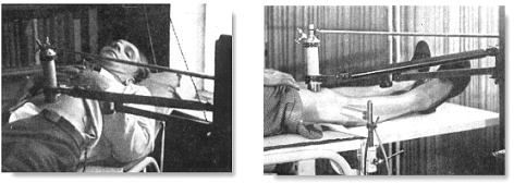
Uses of ultrasonic energy in the 1940s. Left, in gastric ulcers. Right, in arthritis
The 1940s saw exuberant claims made in some sectors on the effectiveness of ultrasound as an almost "cure-all" remedy, abeit the lack of much scientific evidence. This included conditons such as arthritic pains, gastric ulcers, eczema, asthma, thyrotoxicosis, haemorrhoids, urinary incontinence, elephanthiasis and even angina pectoris! Cynicism and concern over harmful tissue damaging effects of ultrasound were also mounting, which had curtailing consequences on the development of diagnostic ultrasound in the years that followed.
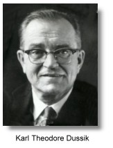 It was around similar times that ultrasound was used experimentally as a possible diagnostic tool in medicine. H Gohr and Th. Wedekind at the Medical University of Koln in Germany in 1940 presented in their paper Der Ultraschall in der Medizin the possibility of ultrasonic diagnosis basing on echo-reflection methods similar to that used in metal flaw detection. They suggested that the method would be able to detect tumours, exudates or abscesses. However they were unable to publish convincing results from their experiments. Karl Theo Dussik, a neurologist/ psychiatrist at the University of Vienna, Austria, who had begun experiments in the late 1930s, was generallly regarded as the first physician to have employed ultrasound in medical diagnosis.
Dussik, together with his brother Friederich, a physicist, attempted to locate brain tumors and the cerebral ventricles by measuring the transmission of ultrasound beam through the skull. Dussik presented his initial experiments in a paper in 1942 and further results after the end of the second world war in 1947. They called their procedure "hyperphonography".
 They used a through-transmission technique with two transducers placed on either side of the head, and producing what they called "ventriculograms", or echo images of the ventricles of the brain. Pulses of 1/10th scond were produced at 1.2 MHz. Coupling was obtained by immersing the upper part of the patient's head and both transducers in a water bath and the variations in the amount of ultrasonic power passing between the transducers was recorded photographically on heat-sensitive paper as light spots (not on a cathode-ray screen). It was an earliest attempt at the concept of 'scanning' a human organ. Although their apparatus appeared elaborate with the transducers mounted on poles and railings, the images produced were very rudimentary 2-dimensional rows of mosaic light intensity points. They had also reasoned that if imaging the ventricles was possible, then the technique was also feasible for detecting brain tumors and low-intensity ultrasonic waves could be used to visualize the interior of the human body.
Nevertheless, the images that Dussik produced were later thought to be artifactual by W Güttner and others at the Siemens Laboratory, Erlangen, Germany in 1952 and researchers at the M.I.T. (see below), as it had become apparent from further experiments that the reflections within the skull and attenuation patterns produced by the skull were contributing to the attenuation pattern which Dussik had originally thought represented changes in acoustic transmissions through the cerebral ventricles in the brain. Research basing on a similar transmission technique was not further pursued, both by Dussik, or at the M. I. T.. For more information read Dussik.
In nearby Germany, Heinrich Netheler, a physician at the Luebeck-South Hospital in Hamburg, was operating in 1945 a small repair facility for medical equipments at the Hamburg university hospital at Eppendorf and had a mission of developing inventive medical products. Professor Hansen, his superior, suggested to him in that year to develop an ultrasonic tomographic equipment for medical use basing on the concept of the RADAR. Important pioneering reseach work started at the Eppendorf University Hospital. Nevertheless, due to a lack of funds right after the war, the equipment designs had not reached the stage of actual fabrication. In the mid 1940s, German physician Wolf-Dieter Keidel at the Physikalisch-Medizinischen Laboratorium at the University of Erlangen, Germany, also studied the possibility of using ultrasound as a medical diagnostic tool, mainly on cardiac and thoracic measurements. Having discussed with researchers at Siemens, he conducted his experiments using the transmission technique with ultrasound at 60 KHz, and rejected the pulse-reflection method. He was only able to make satisfactory recordings of intensity variations in relation to cardiac pulsations. He envisaged much more difficulties would be encounterd with the reflection method. In the First Congress of Ultrasound in Medicine held in Erlangen, Germany in May, 1948, Dussik and Keidel presented their papers on ultrasound employed in medical diagnosis. These were the only two papers that discussed ultrasound as a diagnostic tool. The other papers were all on its therapeutic use.
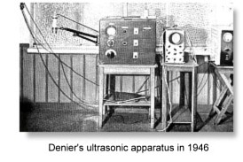 In France, French scientists who were in the study of ultrasonics, namely Andri?Dognon and Andri?Dénier and several others at the research center in Salpêtrière in Paris also embarked on ultrasound insonation experiments before the 1950s. Dénier published his theoretically work on ultrasound transmission in 1946, among many other works on ultrasound used in therapy, and suggested the possibiity of "Ultrasonoscopie". This was a transmission technique and recordings made on a micro-ampere meter and oscilloscope. Equipments were fabricated from 'therapy' counterparts and various electrical current values were determined on different body tissues. Attempts to display voltages as Lissajous figures on the oscilloscope were made. However the work was unsuccessful in producing useful structural images and related instruments were not constructed. Andr?Dénier published in 1951 his book, "Les Ultras-sons -- Appliques a la Medecin". Nearly the entire book was devoted to ultrasonics used in the treatment of various diseases and only a small portion of the text was on ultrasound diagnostics.
 Systematic investigations into using ultrasound as a diagnostic tool finally took off in the United States in the late 1940s. The time was apparently ripe for this to happen. The concept of applying ultrasonics to medicine had progressively matured, so were the available equipments and electronics after the war. George Ludwig, a graduate from the University of Pennsylvania in 1946 was on active duty as junior Lieutenant at the Naval Medical Research Institute in Bethseda, Maryland. There, he began experiments on animal tissues using A-mode industrial flaw-detector equipment. Ludwig designed experiments to detect the presence and position of foreign bodies in animal tissues and in particular to localise gallstones, using reflective pulse-echo ultrasound methodology similar to that of the radar and sonar in the detection of foreign boats and flying objects. A substantial portion of Ludwig's work was considered classified information by the Navy and was not published in medical journals. Although Ludwig's work had started at a considerably earlier date, notice of his work was not released to the public domain until October 1949 by the United States Department of Defence. The June '49 report is considered the first report of its kind on the diagnostic use of ultrasound from the United States.
Ludwig systematically explored physical characteristics of ultrasound in various tissues, including beef and organs from dogs and hogs. To address the issue of detecting gallstones in the human body, he studied the acoustic impedance of various types of gallstones and of other tissues such as muscle and fat in the human body, employing different ultrasonic methodologies and frequencies. His collaborators included Francis Struthers and Horace Trent, physicists at the Naval Research Laboratory, and Ivan Greenwood, engineer from the General Precision Laboratories, New York, and the Department of Research Surgery, University of Pennsylvania. Ludwig also investigated the detection of gallstones (outside of the human body) using ultrasound, the stones being first embedded in pieces of animal muscle. Very short pulses of ultrasound at a repetition rate of 60 times per second were employed using a combined transmitter/ receiver transducer. Echo signals from the reflected soundwaves were recorded on the oscilloscope screen. Ludwig was able to detect distinct ultrasonic signals corresponding to the gallstones. He reported that echo patterns could sometimes be confusing, and multiple reflections from soft tissues could make test results difficult to interpret. Ludwig also studied transmission through living human extremities, to measure acoustic impedance in muscle. These investigations also explored issues of attenuation of ultrasound energy in tissues, impedance mismatch between various tissues and related reflection coefficients, and the optimal sound wave frequency for a diagnostic instrument to achieve adequate penetration of tissues and resolution, without incurring tissue damage. These studies had helped to build the scientific foundation for the clinical use of ultrasound.
 In the following year, Greenwood and General Precision Laboratories made available commercially the "Ultrasonic Locator" which Ludwig used for "use in Medicine and Biology". Suggested usage indicated in the sales information leaflet already included detection of heart motion, blood vessels, kidney stones and glass particles in the body. Ludwig's pulse-reflection methodology and equipment in his later experiments on sound transmission in animal tissues were after earlier designs from the work of John Pellam and John Galt in 1946 at the Electronics and Acoustics research laboratories of the Massachusetts Institute of Technology (M. I. T.), which was on the measurement of ultrasonic transmission through liquids. The M. I. T. was then very much at the forefront of electronics and ultrasonics research. A significant amount of physical data and instrumentation electronics were already in place in the second half of the 1940s, on the characteristics of ultrasound propagation in solids and liquids.
Among other important original findings, Ludwig reported the velocity of sound transmission in animal soft tissues was determined to be between 1490 and 1610 meters per second, with a mean value of 1540 m/sec. This is a value that is still in use today. He also determined that the optimal scanning frequency of the ultrasound transducer was between 1 and 2.5 MHz. His team also showed that the speed of ultrasound and acoustic impedance values of high water-content tissues do not differ greatly from those of water, and that measurements from different directions did not contribute greatly to these parameters.
Ludwig went on to collaborate with the Bioacoustics laboratory at the M. I. T.. His work with physicist Richard Bolt (who, at the age of 34 was appointed Director of a newly conceived Acoustics Laboratory at M. I. T.), neurosurgeon H Thomas Ballantine Jr. and research physicist Theodor Hueter from Siemens, Germany were considered very important seminal work on ultrasound propagation characteristics in mammalian tissues.
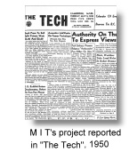
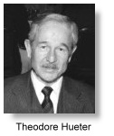 Prior to 1949, Hueter had already been involved at Siemens, Erlangen, Germany, in ultrasonic propagation experiments in animal tissues using ultrasound at frequencies of about 1 MHz, and in ultrasonic dosimetry measurements. These were started in the early 1940s by Ultrasonics pioneer Reimar Pohlman in the same laboratory. In 1948, Hueter met Bolt and Ballantine at an ultrasonic trade show in New York and agreed to join them for new research into the application of ultrasonics in human diagnosis. After a visit to Dussik's department in Austria with Bolt and Ballantine, the group launched a formal project at M. I. T. to perform experiments in through transmission similar to that of Dussik's. Their initial experiments produced results similar to that of Dussik's, and their conclusions were published in their papers in 1950 and 1951 in the Journal of the Acoustical Society of America, and Science. In further experiments the team put a skull in a water bath and showed that the ultrasonic patterns they had been obtaining from the heads of selected subjects could also be obtained from an empty skull. They noted that ultrasonic mapping of the brain tissues within the human skull was prone to great error due to the large bone mass encountered. Efforts were made to compensate for the bone effects by using different frequencies and circuitries, but were only marginally successful at that stage of computational technology.
The M. I. T. research project was subsequently terminated in 1954. They wrote in their paper: "It is concluded that though compensated ultrasonograms (sound shadow pictures) may contain some information on brain structure, their are too sharply "noise" limited to be of unqualified clinical value". The findings had prompted the United States Atomic Energy Commission to conclude that ultrasound will not be useful in the diagnosis of brain pathologies. Medical research in this area was somewhat curtailed for the several years that followed, and enthusiasm was dampened at the Siemens laboratories in Germany to carry out further developments in imaging with ultrasound. At M .I. T. nevertheless, in the course of these pursuits, much basic data essential for tissue characterization and dosimetry were assembled and proved useful for later diagnostic work on other body regions. They had also demonstrated very importantly that interpretable 2-dimensional images was not impossible to obtain. These efforts had paved the way for the subsequent development of 2-D ultrasonic image formation. M. I. T.'s research had also benefited from interactions between the various groups at Champaign-Urbana, Minnesota and Denver.
By the mid 1950s, bibliographic listing of work on ultrasonic physics and engineering applications had totalled more than 6,000. Ultrasonics was already extensively deployed in non-destructive testing, spot welding, drilling, gas analysis, aerosol agglomeration, shear processing, clothes washing, laundering, degreasing, sterilization and, to a lesser extent, medical therapy. Hueter and Bolt's book "SONICS - techniques for the use of sound and ultrasound in engineering and science" published in 1954 became, for example, one of the important treatises in ultrasonic engineering.
 In 1956, D Goldman and Hueter pulled together all the then available data on ultrasonic propagation in mammalian tissues for publication in the Journal of the Acoustical Society of America. The earliest journal devoted entirely to the application of ultrasonics in medicine was "Der Ultraschall in der Medizin" published in Germany. Articles prior to 1952 were entirely on aspects of ultrasound used in therapy. Much of the academic activity at M. I. T. were published in the M. I. T. quarterly progress reports and the Journal of the Acoustic Society of America. After the mid-1950s, due to its ineffectiveness, the transmission technique in ultrasonic diagnosis was abandoned from medical ultrasound research worldwide except for some centers in Japan, being replaced by the reflection technique which had received much attention in a number of pioneering centers throughout Europe, Japan and the United States.
Smaller and better transducers were being assembled from the newer piezoceramics barium titanate after the mid 1940s. They were replaced by lead zirconate-titanate (PZT) when it was discovered in 1954. PZT had a high electro-mechanical coupling factor and more superior frequency-temperature characteristics. The newer transducers had better overall sensitivity, frequency handling, coupling efficiency and output. The availability of very high input impedance amplifiers built from improved quality electrometer tubes in the early 1950s had also enabled engineers to greatly amplify their signals to improve sensitivity and stability.
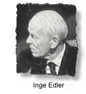 The 'newer' uni-directional pulse-echo A-mode devices developed from the reflectoscope/ metal flaw detectors were soon employed in experiments on medical diagnosis by bold and visionary pioneers around the world. Such were the cases with Douglas Gordon, JC Turner and Val Mayneord in London, Lars Leksell (in 1950), Stigg Jeppson and Brita Lithander in Sweden, Marinus de Vlieger in Rotterdam and Kenji Tanaka and Toshio Wagai in Japan for their pioneering work in the examination of brain lesions. These devices were also employed by Inge Edler and Carl Hellmuth Hertz in Lund in cardiac investigations in 1953, and followed on by Sven Effert in Germany in 1956, Claude Joyner and John Reid at the University of Pennsylvania in 1957 and Chih-Chang Hsu in China, designing their own A- and later on M-mode equipment. Similarily A-mode devices were used in ophthalmologic investigations by Henry Mundt Jr and William Hughes at the University of Illinois in 1956, Arvo Oksala in Finland in 1957 and Gilbert Baum and Ivan Greeenwood in 1955. These uses were all in the 1950s and largely predated clinical applications in the abdomen and pelvis. Researchers in Japan were also actively investigating and producing similar ultrasonic devices and their diagnostic use in neurology, but their findings have only been sparsely documented in the English literature (see below).
John Julian Wild, an English surgeon and graduate of the Cambridge University in England, immigrated to the United States after World War II ended in 1945. He took up a position at the Medico Technological Research Institute of Minnesota and started his investigations with ultrasound waves on the thickness of the bowel wall in various surgical conditions, such as paralytic ileus and obstruction. Working with Donald Neal, an engineer, Wild published their work in 1950 on uni-directional A-mode ultrasound investigations into the thickness of surgical intestinal material and later on the properties of gastric malignancies. They noted that malignant tissue was more echogenic than benign tissue and the former could be diagnosed from their density and failure to contract and relax. Wild's original vision of the application of ultrasound in medical diagnosis was more of a method of tissue diagnosis from the intensity and characteristics of different returning echos rather than as an imaging technique. Between 1950 and 51, he also collaborated with Lyle French at the department of Neurosugery in making diagnosis of brain tumors using ultrasound, although they had not found the method to be very helpful.
 Donald Neal was soon deployed to regular naval services at the naval air base after the Korean war. John Reid, a newly graduating electrical engineer, was engaged through a grant from the National Cancer Institute as the sole engineer to build and operate Wild's ultrasonic apparatus. The device which they first used was an ultrasonic instrument which had been designed by the U.S. Navy for training pilots in the use of the radar, with which it was possible to practise 'flying' over a tank of water covering a small scale map of enemy territory. " We have a tissue radar machine scaled to inches instead of miles by the use of ultrasound". Wild and Reid soon built a linear hand-held B-mode instrument, a formidable technical task In those days, and were able to visualise tumours by sweeping from side to side through breast lumps. The instrument operated at a frequency of 15 megahertz. In 1952 they published the Landmark paper: "Application of Echo-Ranging Techniques to the Determination of Structure of Biological Tissues".
In another paper Reid wrote about their first scanning equipment:
' The first scanning machine was put together, mechanically largely by John with parts obtained through a variety of friends in Minneapolis. I was able to modify a standard test oscilloscope plug-in board. We were able to make our system work, make the first scanning records in the clinic, and mail a paper off to Science Magazine within the lapsed time of perhaps ten days. This contribution was accepted in early 1952 and became the first publication ( to my knowledge ) on intensity-modulated cross-section ultrasound imaging. It appeared even before Douglass Howry's paper from his considerably more elaborate system at the end of the same year.'
 In May 1953 they produced real-time images at 15 megahertz of cancerous growths of the breast. They had also coined their method 'echography' and 'echometry', suggesting the quantitative nature of the investigation. By 1956, Wild and Reid had examined 117 cases of breast pathology with their linear real-time B-mode instrument and had started work on colon tumour diagnosis and detection. Analysis of the breast series showed promising results for pre-operative diagnosis. Malignant infiltration of tissues surrounding breast tumours could also be resolved.
Wild and Reid had also invented and described the use of A-mode trans-vaginal and trans-rectal scanning transducers in 1955. Despite these, Wild was not commended for his unconventional research methods at the time. His results were considered difficult to interpret and lacked overall stability. Intellectual and financial support for Wild's research dwindled, and legal disputes and politics also hampered further governmental grants. His work was eventually supported only by private funds which ran scarce and his data apparently received much less recognition than they deserved.
John Reid completed his MS thesis in 1957 on focusing radiators. In addition he had importantly verified that dynamic focusing was practical. After leaving Wild's laboratory he pursued his doctoral degree at the University of Pennsylvania. From 1957-1965 he worked on echocardiography, producing and using the first such system in the United States, with cardiologist Claude Joyner.
 Visit John Wild's own site on his discoveries and current activities.
 Read also: "The scientific discovery of sonic reflection of soft tissue and application of ultrasound to diagnostic medicine and tumor screening" by John J Wild (Press Release at the Third Meeting of the World Federation for Ultrasound In Medicine and Biology, 1982).
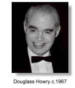
 At the University of Colorado in Denver, Douglass Howry had also started pioneering ultrasonic investigations since 1948. Howry, a radiologist working at the Veteran's Administration Hospital, had concentrated more of his work on the development of B-mode equipment, displaying body structures in a 2-dimensional and sectional manner "comparable to the actual gross sectioning of structures in the pathology laboratory". Published works from the M I T Radar school staff served as initial reference material on techniques in data presentation.
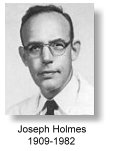
He was able to demonstrate an ultrasonic echo interface between structures or tissues, such as that between fat and muscle, so that the individual structures could be outlined. Supported by his nephrologist friend and colleague Joseph Homles, who was then the acting director of the hospital's Medical Research Laboratories, Howry produced in 1951 with William Roderic Bliss and Gerald J Posakony, both engineers, the 'Immersion tank ultrasound system' *, the first 2-dimensional B-mode (or PPI, plan position indication mode) linear compound scanner. Two dimensional cross-sectional images were published in 1952 and 1953, which convincingly demonstrated that interpretable 2-D images of internal organ structures and pathologies could be obtained with ultrasound. The team produed the formal motorized ' Somascope', a compound circumferential scanner, in 1954. The transducer of the somascope was mounted around the rim of a large metal immersion tank filled with water . The machine was able to make compound scans of an intra-abdominal organ from different angles to produce a more readable picture. The sonographic images were referred to as 'somagrams'. The discovery and apparatus were reported in the Medicine section of the LIFE Magazine® in 1954.
 The 'Pan-scanner' *, where the transducer rotated in a semicircular arc around the patient, was developed in 1957. The patient sat on a modified dental chair strapped against a plastic window of a semicircular pan filled with saline solution, while the transducer rotated through the solution in a semicircular arc. The achievement was commended by the American Medical Association in 1958 at its scientific meeting in San Francisco, and the team's exhibit was awarded a Certificate of Merit by the association.
The work of Douglass Howry, Joseph Holmes and his team is necessarily the most important pioneering work in B-mode ultrasound imaging and contact scanning in the United States that had been the direct precursor of the kind of ultrasound imaging we have today. Pioneering designs in electronic circuitries were also made in conjunction with the development of the B-scan, these included the pulse-echo generator circuitry, the limiter and log amplification circuitry and the demodulator and time gain compensation circuitries.
The Howry/ Holmes systems, although capable of producing 2-D, accurate, reproducible images of the body organs, required the patient to be totally or partially immersed in water, and remained motionless for a length of time. Migration to lighter and more mobile versions of these systems, particularly with smaller water-bag devices or transducers directly in contact and movable on the body surface of patients were imminently necessary.
 Read notes and see more pictures from Gerald Posakony on the early Howry scanners here.
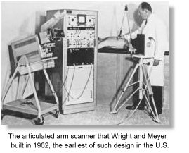
 Homles, together with consultant engineers William Wright and Ralph (Edward) Meyerdirk, and support from the U. S. Public Health Services and the University of Colorado, continued to fabricate a new prototype compound contact scanner, which had the transducer in direct contact with the patient's body and suspended on moving railings above the patient. The apparatus and the usuage of ultrasound scanning were reported in the May 22 issue of the TIME Magazine in 1964.
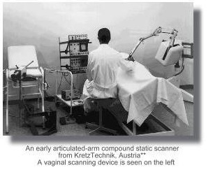
After working on the project for about 2 years, the team finally came up with an innovative multi-joint articulated-arm compound contact scanner with wire mechanisms and electronic position transducing potentiometers. The transducer could be positioned by hand and moved over the scanning area in various directions by the operator.
In 1962, with blessing from Holmes, Wright and Meyerdirk left the University to form the Physionics Engineering® Inc. at Longmont, Colorado, to produce and market their scanner.
 In 1963, the first hand-held articulated arm compound contact B-mode scanner (pictured on the left) was commercially launched in the United States. The launch was reported in the Longmont "Daily Times-Call" in 1963. This was the start of the most popular design in the history of static ultrasound scanners, that of the articulated-arm scanning mechanism.
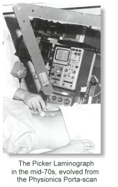
Physionics® was acquired by the Picker Corporation in 1967. Picker continued to produced improved versions of the design right into the 1980s.
Much of the later work in clinical ultrasound was followed up by Homles and his colleagues, Stewart Taylor, Horace Thompson and Kenneth Gottesfeld in Denver. The group published some of the earliest papers in obstetrical and gynecological ultrasound from North America. Douglass Howry had moved to Boston in 1962 where he worked at the Massachussetts General Hospital until he passed away in 1969.
Earliest Wright-Meyerdirk scanner console with one of the first images from a
practical commercial articulated-arm scanner. Portability was also emphasized.
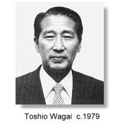 In Japan, at about the same time as Wild and Howry's development, Kenji Tanaka and Toshio Wagai, surgeons at the Juntendo University, Tokyo, together with Shigeru Nakajima, director of the Japan Radio Company, Rokuro Uchida, physicist and chief engineer, had also started looking into the use of ultrasound in the diagnosis of intracranial disease in collaboration with the Nihon Musen Radiation and Medical Electronics Laboratory which had later become the ALOKA® Company in 1950, headed by Uchida. Nakajima and Uchida built Japan's first ultrasonic scanner operating in the A-mode in 1949, modified from a metal-flaw detector. Yoshimitsu Kikuchi, Professor at the Research Institute of Electrical Communications at the Tohoku University in Sendai also assisted in their research. Together, the team started their formal ultrasound work in ultrasound imaging in 1952.
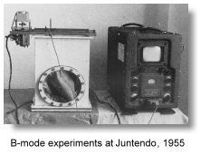 They published 5 papers on ultrasonic diagnosis in brain diseases in that year and many other papers in the ensuing few years. In 1954, Tanaka published an important review entitled "Application of ultrasound to diagnostic field", and investigations had started with other body organs. By 1955, experiments and fabrication with B-mode scanning had started using a similar scope modified from the original A-mode machine coupled with a linear moving transducer gantry. This was shortly developed into the water-bag scanners.
 Also read the Preface and introduction (history) to Tanaka's book "Diagnosis of Brain Disease by Ultrasound" published in 1969 for a short history of his pioneering work in the 1950s.
The M I T hosted a historical conference in Bioacoustics in 1956 and those who attended included Wagai, Kikuchi, Dussik, Bolt, Ballantine, Hueter, Wild, Fry and Howry. Many of them met each other for the first time and important views concerning methods and instrumentations were exchanged at the meeting.
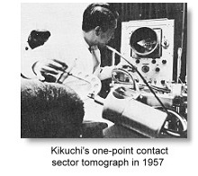 Kikuchi was very active in equipment designs, and by 1957 he was able to demonstrate the "one-point contact-sector scanning tomography" using the plan-position indication (PPI) B-mode format, which had a resemblance to a 'radar display'. This development, which was at around a similar time as the pioneering work of Howry in Denver and Ian Donald in Glasgow (see below), had a similar concept of "position-referenced contact scanning".
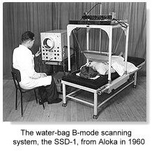
Aloka® produced japan's first commercial medical A-scanner, the SSD-2 and the water-bag B-scanner, the SSD-1 in 1960 (pictured on the right). The application of ultrasound in Obstetrical and Gynaecological diagnosis started around 1956 with the A-scan basing on a vaginal approach and later B-scans at around 1962 basing on the use of the "one-point contact-sector scanner" in the PPI format. Early commercial water-bag scanners were being produced by Aloka® and Toshiba® in the early 1960s.
 Masao Ide at the Musashi Institute of Technology in Toyko, working with Wagai and others launched important pioneering research on the bioeffects of ultrasound. William Fry hosted another conference on ultrasonics in 1962 at the University of Illinois which served as a very important meeting point for researchers from the United States, Europe and Japan.
Michio Ishihara at the National Sanatorium Kiyose Hospital in Tokyo and Hajime Murooka at the department of obstetrics and gynecology, Oomiya Red Cross Hospital, Saitama, delivered the first paper on ultrasound diagnosis of gynecological masses in the Japanese language at the 19th Kanto District Meeting of the Japanese Obstetrical and Gynecological Society in 1958, basing on the A-scan. Murooka had earlier in 1957 received instructions from Wagai on the A-scan methods at the Juntendo University. They described A-scan echoes in cancer of the cervix and also in the presence of different causes of uterine enlargement. Wagai published a review article in the use of ultrasound in Obstetrics and Gynecology in 1959. The Murooka's group apparently did not continue their work after the first two papers presented at scientific meetings.
 Also read a short History of the development of Medical Ultrasonics in Japan.
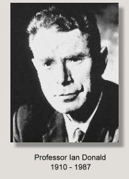
 ohn Wild was back in England in 1954 to give a lecture on his new discovery and this was attended by Val Mayneord, Professor of medical physics at the Royal Cancer Hospital (now the Royal Marsden) who had also been experimenting with the Kelvin & Hughes® MK llB metal flaw-detector in neurological diagnosis. Among the audience was Ian Donald who was then Reader in Obstetrics and Gynaecology at the St. Thomas Hospital Medical School in London and was about to take up the appointment of Regius Chair of Midwifery at Glasgow University. Donald was quick to realize what ultrasound had to offer.# Wild, while returning to Minnesota, had mainly concentrated his investigations on the diagnosis of tumors of the breast and colon using 15 MHz probes which had tissue penetrations of only up to 2 cm. In 1956, Wild published his landmark paper on the study of 117 breast nodules, reporting an accuracy of diagnosis of over 90 percent. Despite that, the ultrasonic method of tissue diagnosis which he so popularised did not reach the point of wide acceptance. Pioneering work in ultrasonic diagnosis in the field of Obstetrics and Gynaecology however, soon took off in Glasgow, Scotland.
The following is an excerpt from an article in the University of Glasgow publication 'Avenue' No. 19: January 1996 entitled ' Medical Ultrasound ---- A Glasgow Development which Swept the World ',
by Dr. James Willocks MD, who had best described the circumstances of Donald's early work :
|
|
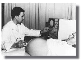
The echoes picked up by the probe are displayed on three oscilloscope screens: an A-scope display, a combined B-scope and PPI display on a long-persistence screen for monitoring: and a similar screen and display of short persistence with a camera mounted in front of it. The probe is moved slowly from one flank, across the abdomen to the other flank being rocked to and fro on its spindle the whole time to scan the deeper tissues from as many angles as possible. ....."
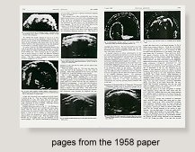
 Ian Donald was also aware of the work of Howry in the United States and Kikuchi in Japan in the early 1950s, and had referenced these pioneers alongside with the work of Wild and Reid in his Lancet paper in 1958. Donald had felt that it was his fortune to have started with these historical A-mode and B-mode instruments instead of the apparatus that Wild and Howry had used, as these involved high frequency transducers (and hence associated with poor penetration into tissues) or a water-bath arrangement which could both become deterrants to further development in a medical setting ##. Aside from this, Donald had on many occasions remarked that a lot of his developments in ultrasound was from a stroke of accident, coincidence and luck. The 'full bladder' was one, which he only discovered in 1963. That the fetal head, being a symmetrical skull bone could be easily demonstrable and measured accurately by a beam of ultrasound in an A-scan was another, as was the opportunity of meeting up with a number of important administrators on the way and working with the very bright engineer Tom Brown from Kelvin & Hughes®.
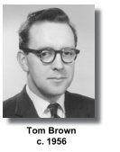
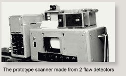
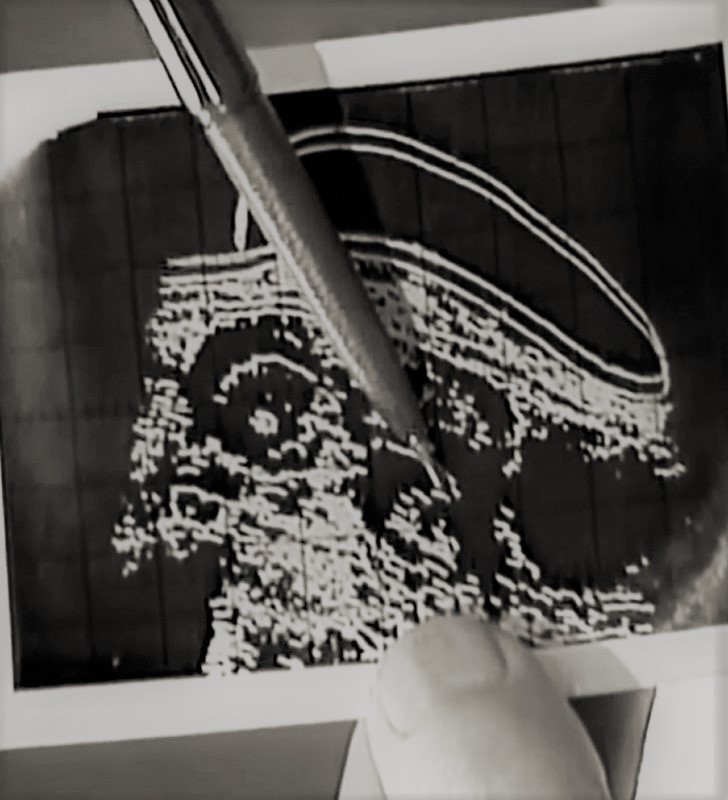
 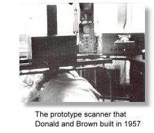 Brown, at the age of 24, invented and constructed with Ian Donald the prototype of the world's first Compound B-mode (plan-position indication, PPI) contact scanner in 1957. The transducer operated at 2.5 MHz. The prototype was progressively improved to become the Diasonograph® manufactured commercially by Smith Industrials of England which had taken control of the Kevin and Hughes Scientific Instrument Company in 1961.
For a detailed account of the pioneering development of the prototypes, read an important unpublished paper by Tom Brown entitled Development of ultrasonic scanning techniques in Scotland 1956-1979.
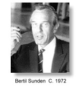
One of Brown's first generation models was sold to Bertil Sunden at Lund, Sweden (see below). The console design of the Diasonograph® came from Dugald Cameron who was then an industrial design student at the Glasgow school of Art. Brown also invented and patented an elaborate and expensive automated compound contact scanner in 1958 and it was at the machine's exhibition in London in 1960 that Ian Donald met for the first time Douglass Howry from the United States who had been using the much larger size water-tank circumferential scanner for several years (see above). Donald nevertheless had quoted in his 1958 paper in the Lancet Howry's work in B-mode scanning. The meeting had also influenced Howry and his team into producing a similar compound contact scanner like the Donald's although this had rapidly evolved into the multi-joint articulated arm version.
A brief description of the working of the prototype compound contact scanner (which eventually developed into the Diasonograph®) was given by Donald and Brown in 1958, the same concept and design were extended into the later commercial models:
" ..... A probe containing both transmitting, and receiving transducers is mounted on a measuring jig, which is placed above the patient's bed. The probe is free to move vertically and horizontally and, as it does so, operates two linear potentiometers, which give voltage outputs proportional to its horizontal and vertical displacements from some reference point. The probe is also free to rotate in the plane of its horizontal and vertical freedom, and transmits its rotation via a linkage to a sine-cosine potentiometer. The voltage outputs from this system of potentiometers control an electrostatic cathode-ray tube, so that the direction of the linear time-base sweep corresponds to the inclination of the probe, and the point of origin of the sweep represents the instantaneous position of the probe. The apparatus is so calibrated that the same reflecting point will repeat itself in exactly the same position on the cathode-ray tube screen from whatsoever angle it is scanned, and likewise a planar interface comes to be represented as a consistent line. 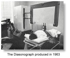


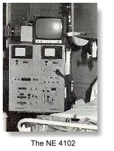
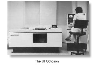
 In 1966, Smiths pulled out of Scotland because the factory was apparently not making money. At the same time the US Supreme Court ruled against Smiths in favour of Automation Industries (formerly the Sperry Company) of Denver on the question of the so-called "Firestone patents" (Floyd Firestone's patent on flaw-detection devices in 1942, see above). As part of the settlement, Smiths undertook to withdraw both from the industrial and medical applications of ultrasound, and Automation acquired title to the collection of Smiths' patents on these subjects. This included the
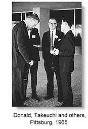






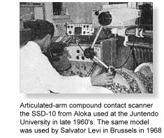 In Japan, Shigemitsu Mizuno, Hisaya Takeuchi, Koh Nakano and Masao Arima followed up the ultrasound work at the Juntendo University in Tokyo, and experimented with new versions of the A-mode transvaginal scanner. The first ultrasound scan of a 6-week gestational sac by vaginal A-scan was reported in the Japanese language in 1963.
From 1962, the group worked extensively with the water-bag B-scanner, the Aloka SSD-1 and was very active in many areas and producing a huge number of research publications, ranging from early pregnancy diagnosis to cephalometry to placentography. They also reported on a large series of pelvic tumors in 1965, and in the following 2 years switched from the water-bag contact scanner to the articulated-arm compound contact scanner, the SSD-10. Another group consisting of T Tanaka, I Suda and S Miyahara started researches into the different stages of pregnancy in 1964.