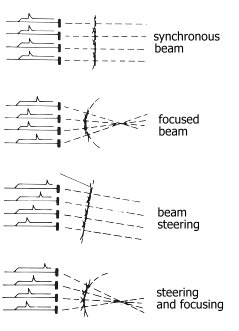
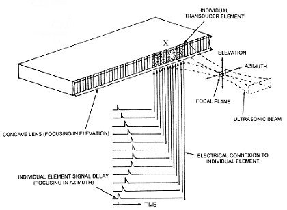
Electronic focusing in the plane along the line of the transducer elements improves lateral resolution as well as sensitivity by increasing the amount of energy in the focal zone. Focusing in the direction at right angles to the scan plane determines the slice thickness and can also be accomplished by use of an external acoustic (light) lens resulting in double enhanced focussing. A unique advantage of electronically focused transducers is the ability to change the depth of the focal zone simply by changing the amount of delays applied to the individual elements. Using pushbutton controls (like in the early ADR), the focal zone can be scanned through a specified range of depths during the real-time exam.
Off-axis beam artifacts were a significant problem in the early linear array designs,. These were the side product of grating lobes which resulted from ultrasound beams that emulated at predictable angles off-axis to the main beam. Grating lobes are unique to array transducers and are caused by the regular, periodic spacing of the small array elements. When the energy of these lobes is reflected by off-axis structures and detected by the transducer, the signal produced is artifactual and produce "ghost images" blurring the main image. To overcome this problems each individual element has been subdivided into a half wavelength wide. This effectively eliminated the grating lobes by increasing the angle to greater than 90 degrees. Eliminating grating lobes also improves the signal-to-noise ratio by increasing the size of the main lobe energy relative to the background energy. This further improves image contrast.
Linear array systems are capable of lateral resolution on the order of less than 1 mm. Axial resolution of 1 mm is always possible depending on the frequency of the system. A "wide aperture" array design means that pulses from a large number (say 128) or all the elements are used to form each scan line. At each line, a different delayed pulse sequencing of the whole array of elements is required to form the unique interference pattern, resulting in a highly focused ultrasound beam perpendicular to the transducer face. Since a unique delay pattern for all the elements is required to produce each scan line, highly sophisticated computer-controlled electronics are required. Lateral resolution of less than 0.5 mm can be achieved.
Annular sector transducer construction varies. All designs sweep the ultrasound beam through a pie-shaped wedge or sector with an opening angle ranging from 30 to 100 degrees. The limited view of superficial structures by sector scanners is offset by their high maneuverability and their ability to visualize large areas at greater depths through small acoustic windows. The mechanical designs enclose the transducer in a fluid-filled case with a flexible membrane that provides acoustic coupling with the skin. The electronically steered-beam, phased-array works in the same principles as the linear sequenced array. Its primary use has been in cardiac imaging where low-amplitude echoes from grating lobe and side lobe artifacts are rejected without loss of significant diagnostic information. However these low-amplitude echoes from diffuse scattering events in soft tissues are essential for imaging in the abdomen and the gravid uterus. Phased-array transducer has not been extensively iused compared with the linear sequenced arrays for general ultrasound scanning.
 The development of 64- and 128-element "steered-beam, phased-array" systems appeared promising for solving the grating lobe artifact problem and improving the signal-to-noise ratio. The electronic complexity is several orders of magnitude greater than that required for conventional linear array systems. All the array elements (64 or 128) must be selectively pulsed to form the wavefront for a single scan Iine. In contrast, only X elements (typically 8 to 16) of the total number of elements (typically 128) are selectively pulsed to form a scan Iine for the common linear designs. Also, the linear array system simply moves the same X elements pulse sequence along the entire array to form parallel focused scan Iines.
The "steered-beam, phased-array" system requires a unique total element pulse sequence for each scan line (typically 128) since each line has its own unique angle with respect to the transducer face in the sector format. The complexity of the newer designs requires sophisticated, high-speed, computer-controlled pulsing of the individual elements circuitry. Electronic focusing on both transmit and receive (similar to annular array designs) provides a longer focal zone with a narrower beam width than conventional single element designs. Similar to linear array designs, focusing in the direction at right angles to the scan plane determines the slice thickness and is accomplished by use of accoustic lens. Since the beam path is electronically controlled, the direction (vector) of each A-line can be selected at random. This unique advantage over mechanical designs allows the system to perform "simultaneous" B-mode imaging and M-mode or Doppler functions.
Back to History of Ultrasound in Obstetrics and Gynecology.

 The development of 64- and 128-element "steered-beam, phased-array" systems appeared promising for solving the grating lobe artifact problem and improving the signal-to-noise ratio. The electronic complexity is several orders of magnitude greater than that required for conventional linear array systems. All the array elements (64 or 128) must be selectively pulsed to form the wavefront for a single scan Iine. In contrast, only X elements (typically 8 to 16) of the total number of elements (typically 128) are selectively pulsed to form a scan Iine for the common linear designs. Also, the linear array system simply moves the same X elements pulse sequence along the entire array to form parallel focused scan Iines.