Historical Notes from Mr. Gerald Posakony:
I first started working with Dr. Douglass Howry in 1949 when he moved to Denver to work with Roderick Bliss in a company called Decimeter, Inc. Rod Bliss had an excellent background in electronics and microwaves and Howry relied on him to help him understand what conceptually could be done in the field of ultrasonics that might be analogous to the field of microwaves and useful to the medical diagnostic field. Howry was an avid reader and his mind was going around constantly. Howry and the team at the Veterans Administration and then at the University of Colorado, Medical Center in Denver, designed, developed and demonstrated four different diagnostic systems. I will discuss the details and limitations........ .
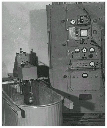
The picture shows the original reduction to practice of Howry's "Somascope". I did most of the mechanical, the acoustic and transducer design as well as some of electronics, but C. R. Cushman did most of the electronics. The picture shows the converted radar frame and the horse trough to which we adapted the rails. Of most significance in this picture is the tube holding the transducer. This was a focused transducer made of lithium sulfate. It had a fairly narrow beam and a broad bandwidth signal response. This gave us high axial and lateral resolution. The transducer head could be in moved linearly back and forth or it could oscillate back in a polar rotation over about 120 degree angle or it could do both. This latter consideration allowed us to see biological structures that where in the field of view of the sound beam.
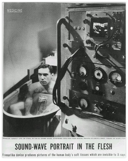
This picture shows the system and operation. I happen to be the subject and my kidney can be seen on the oscilloscope screen. This picture appeared in Life magazine in the medical section in 1954.
The results from a long series of studies clearly showed us that a conventional linear back and forth motion and/or a simple polar oscillation would not provide the kind of detail required for medical diagnosis. It caused us to study the problem in more detail. The conceptual idea was that we should build scanner that could rotate around the patient and the transducer should shuttle back and forth to see all structures in a tubular like (the body) structure. This resulted in a compound scan that allowed the sound beam to see biological tissue regardless of position. In other words we were looking at tubular or cylindrical like body structures, accommodating the shape of the subject. This resulted in the development of the 360 degree water-bath scanner shown here below:
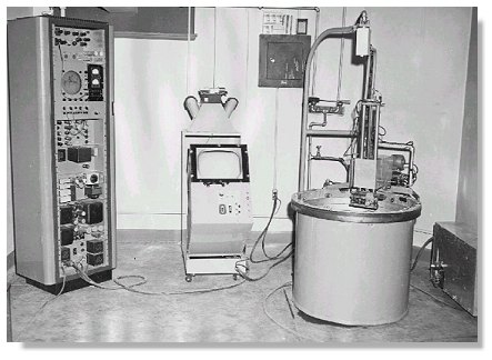
This is the picture of the scanner. The water-bath scanner consisted of a B-29 gun turret, electronics console and display unit. Not only is the transducer scanned a full 360 degrees (or portions thereof) but the transducer itself is shuttled back and forth about 200 mm. The subject was immersed in the tank and a lead weight lied on his abdomen to prevent floating. Doing a geometric deconvolution, we deducted that with this configuration, the sound beam would interrogate the subject from appropriate positions and see structures that were somewhat off the normal incidence seen by conventional back and forth scanners. In those days we did not have the computers to reconstruct our images.
The most dramatic picture and the one demonstrating the concept is the cross-section of the neck of our associate, C. R. Cushman.
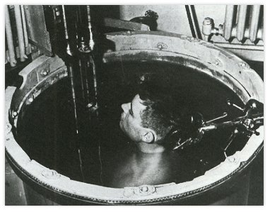
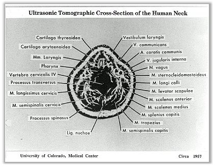
This cross-section picture shows not only the arteries and veins, but more dramatically the vocal cords et al. The rest of the structures are defined in the picture. This picture took several minutes to make and Mr. Cushman sat very still with a lead brick in his lap.
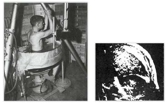
The third system is the Pan Scanner. This design was an attempt to develop a system that did not require full immersion but could still take advantage of a controled sound beam (focus) and compound scanning. It emplyed a semicicular pan with a section removed from its flat surface and had plastic sheeting inserted over the hole. Mineral oil was put on the patient's skin to provide sonic contact when the patient was strapped against the plastic window. A dental chair raised and lowered the patient at the pre-selected distances. The scanning carriage moved in a semicircular path and the transducer moved simultaneously in a horizontal 4-in path. This type of equipment provided good scans of the liver, spleen, kidneys and bladder. It was also possible to obtain excellent breast scans by cupping the plastic sheet and positioning the patient against the sheet.The scan on the right showed the kidney and the liver. We used photographic film. A camera, with an open shutter, recorded the signals on the CRT in term of bright spots on the monitor. It took four or five "hits" from the same internal structure for the "spot" to bright enough to record on the film. While the image could be sen developing on the CRT, one had to wait till the film was developed to see the final product. The team's achievement was commended by the American Medical Association in 1958 at their Scientific meeting at San Francisco, and the team's exhibit was awarded a Certificate of Merit by the Association.

A Certificate of Merit was awarded to the Howry team by the American Medical Association in 1958 at its Scientific meeting in San Francisco, for its acheivement in visualising body tissues by ultrasound.
System No. 4 was a rather large device using a gantry to hold and manipulate the transducer. I do not have a photograph of this system but it appeared in Medical Instrumentation about 1961. In operation, the patient would lie on a table. The transducer was in contact with the patient and was moved across the patient by an operator. The resolvers on the gantry provided the the X, Y and Z positional information. A gel was used to couple the ultrasound to the patient so no immersion was required. The system worked well, but was quite cumberson.
By this time (early 60's) Wright and Meyer were working on the project at the University of Colorado as members of the Howry/Holmes team. They were astute enough to recognize that the concept of moving the transducer and imaging on a storage oscilloscope (which had just come into popular use) could be used to achieve nearly the same results as was being achieved with the big gantry mechanical system, by using a pantograph arm of the type used in mechanical drafting. Outside the University, they started the company Physionics Engineering, which the eventually sold to Picker (as I remenber).
* Notes and images courteously provided by Mr. Gerald Posakony. Reproduced with permission.
Back to History of Ultrasound in Obstetrics and Gynecology.