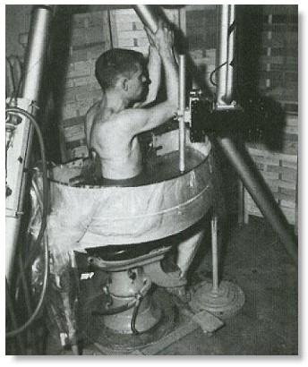
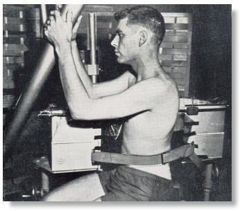
Picture of the Pan Scanner, front and back side. This design was an attempt to develop system that did not require full immersion but could still take advantage of a controlled sound beam (focus) and compound scanning. The tank was semi-circular with plastic film in contact with the skin of the patient. The transducer carriage was mounted on a tripod above the pan and moved in a semicircular path. The patient was held against the pan by straps. The subject was seated on an old dental chair which was moved up and down to obtain serial cross sections. With this equipment excellent studies were obtained on liver, spleen, kidney and bladder.
In those days there was no computer to reconstruct the images. The team used photographic film. A camera, with an open shutter, recorded the signals on the CRT in term of bright spots on the monitor. It took four or five "hits" from the same internal structure for the "spot" to bright enough to record on the film. While the image could be seen developing on the CRT, one had to wait till the film was developed to see the final product.
-- from Mr. Posakony.
Back to History of Ultrasound in Obstetrics and Gynecology.

